Cavitational lesions are difficult to discover. On most x-rays, unless the doctor is specifically trained, these bony lesions are usually missed.
Gross examination of NICO lesions are shown in Figure 2. Note the large nerve, the inferior alveolar, as it travels through and between NICO lesions.
Dental students and residents spend a lot of time learning to properly read x-rays of all types. A very useful x-ray view in dentistry is the panoramic radiograph. Unfortunately, all of us were trained to read certain irregularities as normal! We now know that these irregularities on panoramic x-rays are quite often cavitational lesions.
Figure 3: Gross appearance of NICO lesions

Note that at least 4 lesions are visible. IAN: inferior alveolar nerve.
Figure 4: A normal panoramic x-ray?

See the panoramic x-ray above (Figure 4)? Most dentists, oral surgeons, radiologists, and other doctors would read this x-ray as normal.
Figure 5: NICO lesions in left posterior mandible.

Now, look again at the same x-ray, but with lines drawn to the NICO lesions (Figure 5).
This 44 year old lady had left lower jaw pain for a couple of years, after the last two molars were treated with large fillings, then root canals, and then removed. She also had a slow drainage into her mouth which produced a sore throat. Unfortunately, this lady saw at least 8 doctors (a dentist, 2 oral surgeons, a periodontist, an endondontist, 2 ENT physicians, and a family physician) and all could find nothing and even suggested she consult a psychologist!
Here’s another interesting case. This is a 47 year-old business woman who has had extensive and good dental treatment. Her wisdom teeth were taken out when she was 14 years old and the surgery was difficult. When she was first married at age 18, she took birth control pills for only a few weeks because she developed phlebitis in the deep veins of her legs. When she was 45 or 46, she began experiencing deep aching pain in her lower right jaw. There was no swelling, but she complained of a terrible, sour taste.
Figure 6: An apparently normal panoramic x-ray.
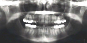
Look at Figure 6. This is her panoramic x-ray. From the looks of this x-ray, there appears to be nothing wrong, yet she had continual deep aching pain in the right lower jaw, a sour taste and no teeth which seemed painful or sensitive.
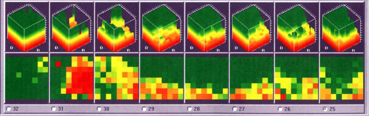
Figure 7 is a copy of this lady’s Cavitat or ultrasonic scan of her lower right jaw. Tooth #28 was removed years earlier for othodontic reasons.
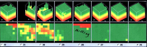
Figure 7: Ultrasound (Cavitat) scan of lower right jaw.
Note the red, yellow and brown colors in the areas of teeth numbers 29 through 32. These colors indicate areas in the bone of reduced blood flow or dryness, or in other words, a cavitation or cavitations. When this lady’s lower jaw was numbed with a local anesthetic, all her pain subsided, but her sour taste persisted. This, along with her symptoms and Cavitat scan, indicated that she had a cavitation in the areas of teeth numbers 29 through 32.
Figure 8: Cavitation in lower right jaw at surgery.

Figure 8 is a picture during surgery of this area. Note the large, void area in the jaw bone. This cavitation area was present within the bone and not created by Dr. Shankland. The two last molars were removed, but the cavitation is lateral to the teeth. Her surgery was difficult and she had minor nerve damage for a few weeks. But today, more than four years after the surgery, she’s pain-free and no longer has a sour taste in her mouth.
Figure 10: Cavitation in upper jaw of 58 year-old woman.

Not to belabor the point, look at Figure 10, which shows yet another case. Is there any doubt that there’s a hole in this lady’s upper jaw? At this point, only the gum tissue was lifted up; no bone was removed. This lady had constant upper jaw pain, pressure and a sour taste in her mouth. She was diagnosed with trigeminal neuralgia and scheduled for brain surgery. This bone lesion went clear through her jaw into her palate and up into the floor of her nose.
Figure 11: Cavitat scan of lower right jaw.
In one last case, this lady had undiagnosed right facial pain for years. She complained of a sour taste at times and when the sour taste wasn’t present, she’d have intense pressure in her right jaw. Figure 11 shows her ultrasound or Cavitat scan. Note the red area in the area of #31 and as it extends into the area of #30. Again, this shows an area of ischemia, or reduced blood flow, which is actually a jaw bone cavitation.
Figure 12: Panoramic x-ray of the same patient shown in Figure 11.
Figure 12 shows a panoramic x-ray of this same patient. If you look closely at the x-ray, it will look normal and you’ll not be able to see any abnormality. Yet, look at Figure 13, a picture taken after an incision was made and nothing else done at the surgical site. Look at the large cavitation at a former extraction site.
Figure 13: A cavitation visible after an incision and the gingival tissue is retracted. This is a former extraction site.

Figure 14: The same cavitation with the bony roof removed.

Now, look at Figure 14, a picture of the same patient with the roof of the cavitation removed for access to surgically repair the area. Isn’t that amazing? Several fine doctors couldn’t diagnose this lady’s problem and most thought she was crazy! Fortunately, she’s doing fine with no further pain, sour taste and pressure and no nerve damage after the surgery.